Location
Get in touch
Call: 308-630-1140
Login to MyChartWhat is a PET/CT exam?
A Positron Emission Tomography (PET) and Computed Tomography (CT) exam combines two scans into one to create images with information about both anatomy and physiology. PET uses an injected radioactive isotope that travels through the blood stream and attaches to different types of cells in the body. These areas of activity may be normal or indicate disease, depending on where they are located. The CT portion of the exam is used to create images that help precisely locate where the areas of activity are within the body. PET/CT exams are used to help treat and diagnose illnesses such as cancer, heart disease, or brain disorders (such as Alzheimer’s disease or dementia).
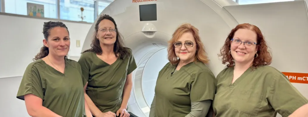
Regional West’s PET/CT suite is located in Regional West Medical Plaza South and features a Siemens Biograph mCT PET/CT machine, offering full diagnostic CT capability, lower radiation doses and exposure, and increased image quality and scan speeds. We are also proud to offer specialty PET/CT exams, such as prostate-specific membrane antigen (PSMA) PET/CT exams, and Cu-64 Detectnet (Dotatate) scans for neuroendocrine tumors.
Your primary care physician or provider must refer you for a PET/CT exam. Since the radioactive isotope used for PET/CT exams is created specifically for each patient and his or her appointment time, we need at least one day’s notice to order the isotope. Because these drugs expire within hours, it is important to be on time for a nuclear medicine exam or let the Imaging Services department know at least one day ahead if you need to cancel or reschedule an appointment. You can call or reschedule by calling the Scheduling Services at 308-630-2700. To reach the Imaging Services main desk, call 308-630-1140.
What to expect during a PET/CT exam
For a PET/CT exam, you will first come to the PET/CT suite for the radioactive isotope injection. The amount of radiation in this is small, so your risk of negative radiation effects is low.
Then, you will rest quietly for approximately 60 minutes so the isotope has time to circulate through the body. Because this study relies on the body’s use of sugar, it is important to keep muscles relaxed right before the exam, so they aren’t using sugar during the scan. You are also asked to restrict your diet and cut out sugar for 24 hours before the exam.
After the relaxation time, you will lie on the table while the CT scan is performed and the computer creates cross-sectional images. While you are on the table, the PET camera will measure the radioactivity coming from within your body. Computers will then create an image of the body that shows the location where the radioactivity collects. Images of the areas of activity are fused into the CT scan images, creating cross sectional images that can show precisely where the areas of radioactivity are located.
During your exam, a registered nuclear medicine technologist will care for you. The technologist is specially trained in performing nuclear medicine exams and certified in this specialty by the American Registry of Radiologic Technologists (ARRT) or the Nuclear Medicine Technology Certification Board (NMTCB). The technologist will inject the radioactive isotope. He or she will assist you onto the scanning table and operate the equipment. The technologist will be with you throughout the exam.
A board certified radiologist who specializes in PET/CT exams will examine the images. Then, they will send the report directly to your physician or provider, who will then contact you with the results.
For more information on specific exams, as well the pre-exam diet plan, check out the fact sheets below.
- Click here to learn more about PSMA PET/CT exams.
- Click here to learn more about skull base to mid-thigh FDG-PET exams.
- Click here to learn more about whole body FDG-PET exams.
- Click here to learn more about basic brain FDG-PET exams.
Click here to see the FDG-PET diet plan.